Ocular Services @ LifeCare Pet Hospital
Also Serving South Riding, Aldie, Ashburn, Centreville, Reston And Herndon
- Complete eye examination and diagnosis
- Treatment planning which may be medical or surgical depending on the condition.
- Surgical equipment includes an operating microscope, and very fine surgical instruments and sutures which allow optimal tissue apposition and minimizes scarring.
- Medical and surgical treatments for eyelid conditions and tumors, entropion, cherry eye, corneal ulcers and microsurgery, dry eye, and more are offered. Lens/cataract surgery is not available.
- Intrascleral prosthetics are available for some cases that require eye removal.
Eye Examination
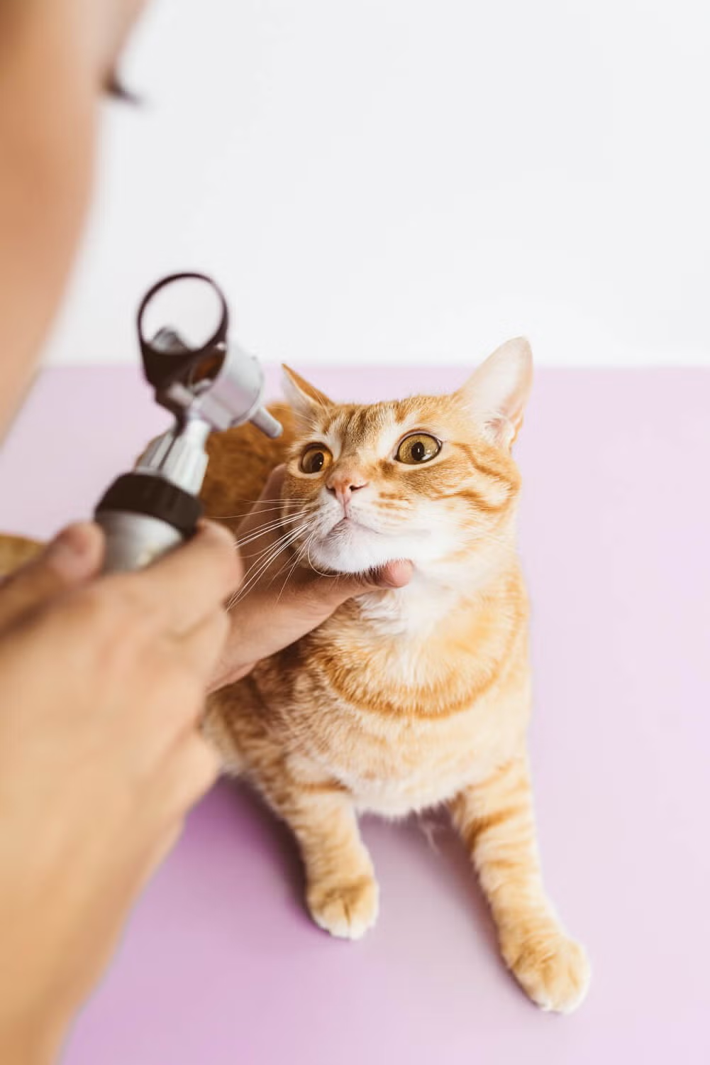
 Your pet’s eye examination will be similar to that performed on a person. However for a full and accurate examination, a dark area will always be needed for assessment of the eyes.
Some of the tests that may be performed during your pet’s assessment include:
Schirmer tear test
A special sterile soft paper strip is placed into the eye to measure the watery tear production over a 60 second period.
Fluorescein stain
This is yellow / green stain used for examining the surface of the eye – the stain attaches to scratches and ulcers on the corneal surface allowing evaluation of the size, severity and progression of the injury. It may also be used to assess the normal tear flow out of the eyes into the nose / mouth.
Rose Bengal stain
This is another examining stain, which attaches to areas of more subtle damage to cornea and conjunctiva.
Biomicroscopy
Or slit lamp biomicroscopy is a valuable examining instrument for highly magnified assessment of the front part of the eye (cornea, iris and lens) and eyelids.
Tonometry
This is the measurement of the pressure inside the eye and is measured with a “Tonometer”. A high pressure indicates glaucoma, a low pressure may indicate inflammation within the eye.
Ophthalmoscopy
This is the visualization and assessment of the back of the eye (the retina / fundus) using an ophthalmoscope.
Medical therapy for ocular conditions @ LifeCare Pet Hospital
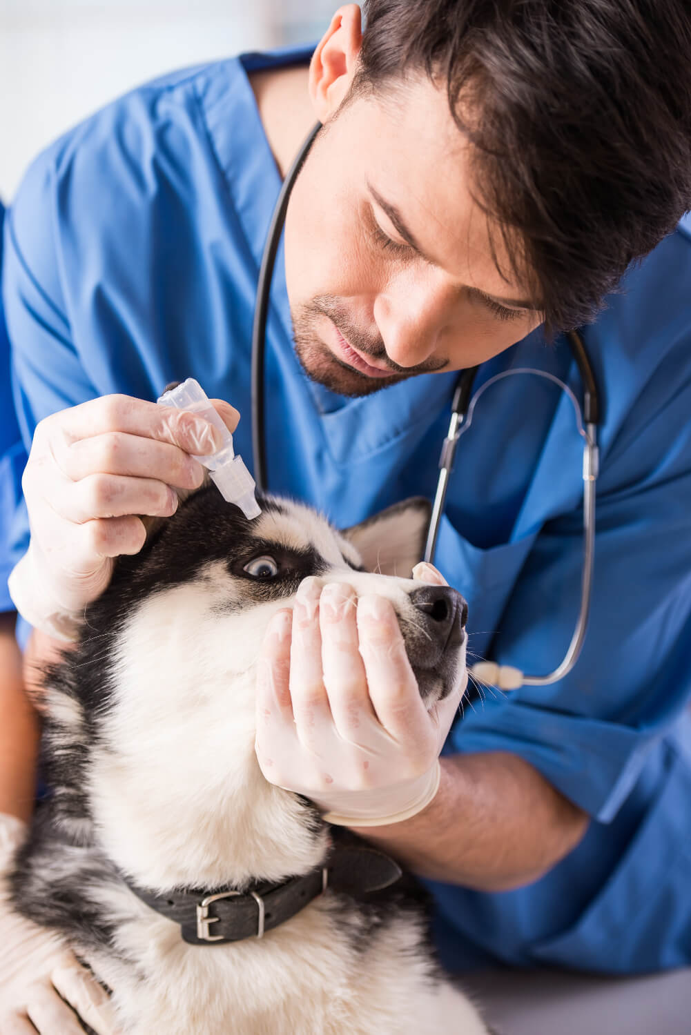
We keep broad range of medications on hand to treat all of your pet’s ophthalmic problems. Our goal is to ensure your pet remains as comfortable as possible during treatment, and to aid in the healing process in any way possible.
Topical Antibiotic Medications
For all-encompassing treatment of the full spectrum and variety of corneal ulcers.
Anti-Glaucoma Medications
For sight-preserving and comfort-maintaining therapy designed to help keep elevated eye pressure under control.
Anti-Inflammatory Medications
For treatment of inflammatory and painful eye diseases such as uveitis, and alleviation of post-surgical discomfort.
Analgesic / Pain Medications
To ensure that your pet always remains as comfortable as possible during treatment of his or her ocular condition.
When Medicine Alone Is Not An Option For Treatment
Surgical therapy for ocular conditions offered at LifeCare Pet Hospital
- Eyelid Tacking
- Ectopic Cilia Excision
- Eyelid Mass Removal
- Eyelid Reconstructive Surgery:
- Hotz-Celsus for Entropion
- Medial Canthoplasty
- Z-plasty
Third Eyelid Surgeries
 Nictitans Gland Replacement (“Cherry Eye” Surgery)
A prolapsed gland is the most common disorder of the third eyelid in dogs. It is commonly referred to as a cherry eye because the prolapsed gland appears as a red mass that protrudes from behind the third eyelid. Prolapsed gland of the third eyelid is more common in young dogs and is overrepresented in some breeds, including American cocker spaniels and English bulldogs.
The exact cause of this condition is unknown, but it is thought to be due to a weakness of the tissues that normally anchor the gland to the periorbita. The condition may be unilateral but is often bilateral, with the glands prolapsing at different times.
Corneal Surgeries
- Keratectomy
- Conjunctival Grafts
- Corneal Laceration Repair
Eye Removal Procedures
The operation involves a general anaesthetic for your pet. The hair around the eye is clipped; the eye is completely removed and the eyelid skin is then stitched closed over the eye socket (the bony space within the skull that protects the eye). Stitches, which may require removal, may or may not be placed within the outer layers of the eyelid skin. Pets will be able to go home on the day of surgery to recover comfortably in their home environment.
At LifeCare Pet Hospital, we’re committed to helping your pet move comfortably and live their best life. With our compassionate care and expertise, your furry friend is in good hands.
